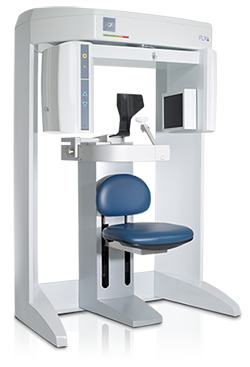
 While CT (computed tomography) imaging has been used in the medical field for over 30 years, it is becoming the new diagnostic tool of choice for orthodontic analysis, diagnosis and treatment planning due to the latest advancements in diagnostic technology such as the revolutionary i-CAT® Cone Beam 3-D Imaging System. This three-dimensional CT technology can provide a quicker full scan of the head than traditional two-dimensional imaging, allowing orthodontists a better visualization of the hard and soft tissues of the craniofacial structures from several perspectives. In addition to the advantages of traditional CT scans, the i-CAT® Cone Beam releases up to 90% less radiation than traditional X-ray machines, enhancing your safety while providing crisp, clear images for more efficient diagnostic analysis and treatment. We feel our modern, cutting-edge techniques ensure you are receiving the quality care you deserve.
The i-CAT® Cone Beam 3-D Imaging System can immediately produce three-dimensional images in under one minute. This in-office, easy-to-use system provides your orthodontist a comprehensive view of all oral and maxillofacial structures, dramatically increasing the efficiency with which your orthodontist is able diagnose your condition and plan for your treatment.
i-CAT® technology is used for:
- TMJ assessment
- Surgical planning
- Assessment of cleft lip and palates
- Assessment of the alveolar bone
- Impacted tooth position
- Facial analysis
- Tongue size and posture
- Airway assessment
- Placement of dental implants
